In vivo antioxidant potential of lepidium sativum l. Seeds in albino rats using cisplatin induced nephrotoxicity
Keywords:
Cisplatin, Nephrotoxicity, urea, creatinine, glutathione, Lipid peroxidationAbstract
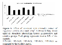
The present study was designed to investigate to possible potential nephrocurative, nephroprotective activity and in vivo antioxidant potential of 200mg/kg and 400mg/kg ethanolic extract of Lepidium sativum L. seeds was use to against cisplatin (5mg/kg, i.p.) induced nephrotoxicity. The experimental protocol designed as the animals were divided into six groups (n=6) like control, model control, two curative (200mg/kg and 400mg/kg), and two protective groups (200mg/kg and 400mg/kg, were received vehicle, cisplatin, cisplatin + extract, and extract + cisplatin respectively. After 6th days, blood collected from retro-orbital sinus of rats and determined urea and creatinine level in serum of each group after then rats were sacrificed for quantitative estimation of various enzymes and ATPase content in kidney tissue. A single dose of cisplatin induced loss in body weight, increase urine excretion, increased urea & creatinine level in serum; it was significantly recovered by 200mg/kg and 400mg/kg in curative and protective groups. The enzyme estimation in kidney tissue it found that increase malondialdehyde, superoxide dismutase, catalase and reduced glutathione level, it was significantly monitored by 200mg/kg and 400mg/kg in curative and protective groups. These are defined as vivo antioxidant potential. The level of brush border enzymes like Na+ / K+ ATPase, Ca++ ATPase and Mg++ATPase were found significantly reduced after single dose cisplatin injection. It was overcome by treatment of same extract in curative and protective groups. Finally it is concluded that the present study data conformed nephrotoxicity induced by cisplatin due oxidative stress and ethanolic extract of Lepidium sativum L. seeds may have nephroprotective and curative activity.
References
Bergstrom J, Furst P, Norée LO, Vinnars E.
Intracellular free amino acid concentration in
human muscle tissue. J Appl Physiol 1974;
; 693–6.
Nadkarni′S K.M. Indian materica medica.
Reprint 1995; I; 736.
Welbourne TC. Ammonia production and
glutamine incorporation into glutathione in
the functioning rat kidney. Can J Biochem
; 57:233–7.
Kirtkar K M, and Basu BD. Indian medicinal
plants. 2005; I: 174.
Gonzalez Ricardo, Romay Cheyla, Borrego
A, Merino FH, Zamora MZ, and Rojas E.
Lipid peroxides and antioxidant enzyme in
cisplatin induced chronic nephrotoxicity in
rats. Mediators Inflamm. 2005; 3: 139-143.
Kersten L, Braunlich H, Kepper BK, Gliesing
C, Wendelin M, Westphal J. Comparatively
Antitumoral - active platinum and ruthenium
complexes in rats. J appl toxicol 1998; 18(2).
Zhang JG, Lindup WE. Role of mitochondria
in cisplatin - induced oxidative damage exl
slices. Biochem pharmacol 1993; 45 (11); 677
– 683.
Kaplan A, Lavemen IS. The tests of renal
function, in clinical chemistry. Interpretation
and techniques 2nd ed., lea and Febiger,
Philadelphia, 1983; 109-142.
Varley, and Alan HG. Tests in renal disease.
In Practical Clinical Biochemistry. 1984; 1:
Sedlak M, and Lindsay R. Estimation of total,
protein-bound and non-protein sulfhydryl
groups in tissue with Ellman’s reagent. Anal
Biochem; 1968; 25:192–205.
Hartree E. Determination of protein: a
modification of long method that gives a
linear photometric response. Anal Biochem
; 48: 4227.
Didier Portilla, Richard TP, Safar AM.
Cisplatin-induced nephrotoxicity Last
literature review version. 2008; 16.3.
Uchiyama M, Mihara M. Determination of
malonaldeyde precursor in tissues by
thiobarbuturic acid test. Anal Biochem; 1978;
: 271–8.
Mishra HP, Fridovich I. The role superoxide
anion in the auto-oxidation of epinephrine
and a simple assay of SOD. J Bio chem1972;
: 3170.
Sinha A. K., colorimetric assay of catalase,
anal biochem, 1972, 47,389.
Bontin SL, Presence of enzyme system of
mammalian tissue. Wiley inter sci.
,197,257.
Hjerken S, Pan H. Purification and
characterization of two form of the low
affinity calcium ion ATPase from erythrocyte
membrance. Biochem Biophys Acta,
,728,281.
Ohinishi T, Suzuki Y, Ozawa K. Comparative
study of plasma membrance magnesium ion
ATPase activities in normal regenerating and
malignant cell. Biochem biophys ACTA,
, 684, 64.
Mora l deo antune, LM. Francescato HD,
Bianchi M del, the effect of oral glutamine on
cisplatin –induced nephrotoxicity in rats.
pharmacol.Res. 2003; 47, 517-522.
Matsushima H, Yonemura K, Ohishi K,
Hishida A. The role of oxygen free radicals in
cisplatin-induced acute renal failure in rats. J
Lab Clin Med, 1, 1998; 31:518–26.
Daugaard G, Abilgaard U, Holstein-Rathlou
NH, Bruunshuus I, Bucher D, Leyssac PP.
Renal tubular function in patients treated with
high-dose cisplatin. Clin Pharmacol Ther
; 44:164–72.
Naziroglu M, Karaoglu A, Aksoy AO,
Selenium and higher dose vitamin E
administration protects cisplatin induced
oxidative damage of renal, liver, lens, tissue
in rats. Toxicolo, 2004; 195: 221-239.
Francescato HDC, Costa RS, Camargo SMR,
Zanetti MA, Lavrador MA, Bianchi MLP.
Effect of dietary oral selenium on cisplatin
induced nephrotoxicity in rats. Pharmacol Res
; 43:77–82.
Antunes LMG, Darin JDC, Bianchi MLP.
Effects of the antioxidants curcumin or
selenium on cisplatin-induced nephrotoxicity
and lipid peroxidation in rats. Pharmacol Res
; 43:145–50.
Nelson DL, and Michael M COX. Lehninger
principle of biochemistry. 4th edition 2005;
-858.
Molitoris BA, Dahl R, Geerdes A.
Cytoskeleton distribution and apical
redistribution of proximal tubule Na+/K+
ATPase during ischemia, Am J. Physical,
,265,F488-F495.


